Secrets of cell division revealed by cutting-edge imaging technique
Research from University of Copenhagen Associate Professor Fena Ochs enhances our understanding of cell division by identifying distinct cohesin populations within cells. The study utilized super-resolution microscopy to visualize the protein complexes at the molecular level.

An innovative study by Fena Ochs, new Group Leader and Associate Professor at Biotech Research & Innovation Center (BRIC) University of Copenhagen, delves deep into the intricate world of cell division. The study, published in Science, sheds light on the role of cohesin, which is a crucial protein complex that helps to faithfully segregate genetic material during cell division.
Cell division is the cornerstone of life, transforming a single cell into the complex organisms we see around us. Yet, the process itself is a delicate dance of molecular interactions, particularly the task of dividing genetic material accurately between two daughter cells, then into 4, 8, 16 cells and so on. The intricate choreography is orchestrated by cohesin, a protein complex that forms a ring holding identical chromosomes together until the moment of division.
Using cutting-edge super-resolution microscopy, the research team zoomed into human cells to visualize cohesin complexes at an unprecedented level of detail. What they discovered was remarkable: distinct populations of cohesin complexes, each playing a specific role in our cells, explains corresponding author Associate Professor Fena Ochs,
“This breakthrough not only advances our fundamental understanding of cell division but also holds promise for many other research fields. By showcasing the power of single-molecule super-resolution imaging, this study opens new doors for studying biological processes in their natural environment with minimal disturbance,” she says.
Specifically, the study revealed the existence of multiple populations of cohesin complexes within cells. One of the cohesion populations discovered is the one responsible for sister chromatid cohesion, the process critical for generation of healthy new cells during cell division. The second population includes cohesin responsible for loop extrusion. Loop extrusion is the process of compacting our DNA to fit into our cells and to promote important interactions between genetic elements.
Amazing level of detail in microscopic cellular processes
The technique utilized by the research team is based on a type of super-resolution microscopy called Structured Illumination Microscopy, which the researchers now have further developed for quantitative imaging at the single molecule level. Normally, researchers break apart cells to be able to study the processes within them, says Fena Ochs.
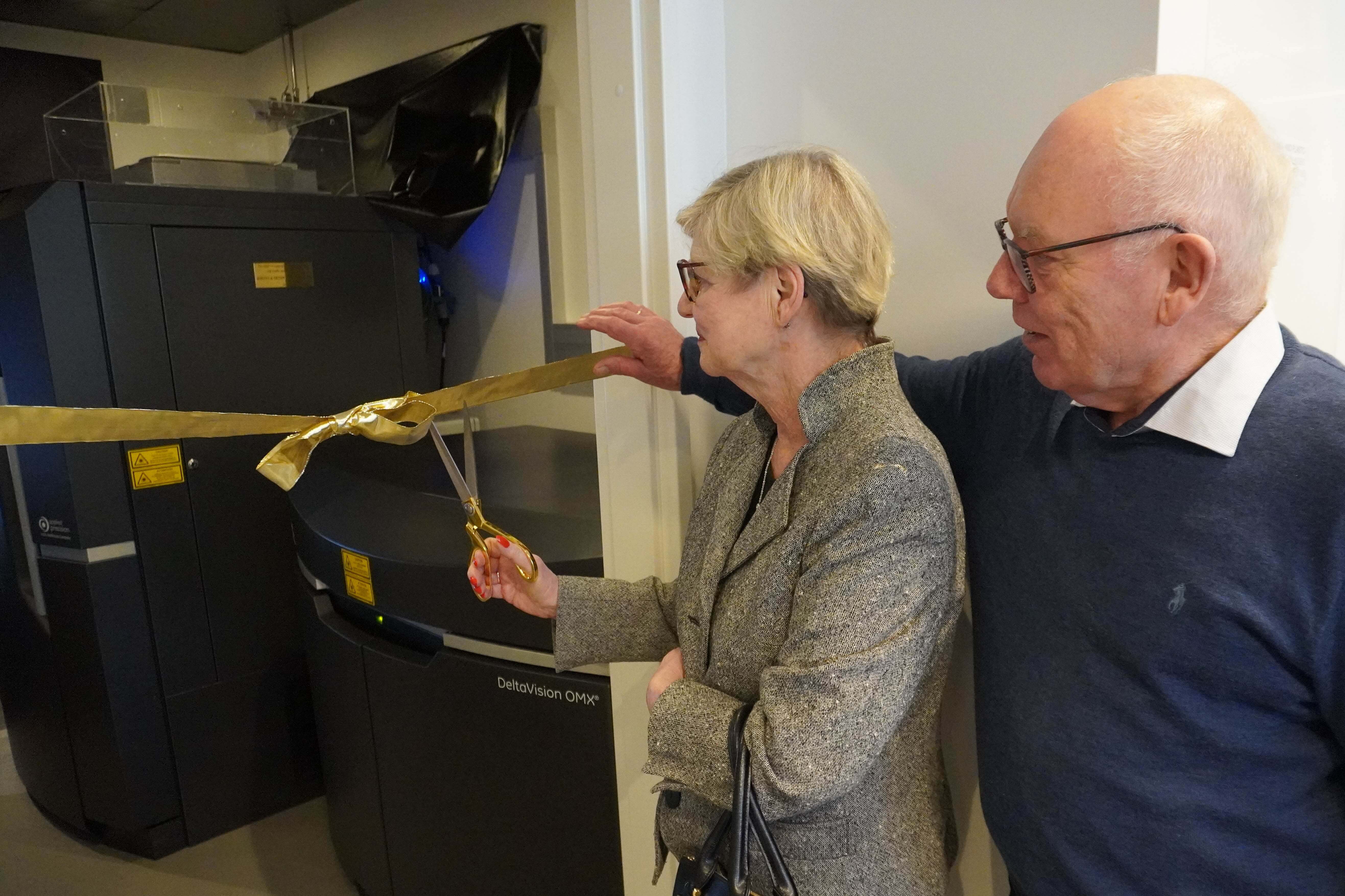
“With our new microscopy approach, we now have the opportunity to image molecular processes in intact cells at incredibly high resolution and without disturbing them much. We've just acquired the new microscope, so this is only the beginning.”

While this study was carried out at the University of Oxford, where the technology was invented, Fena Ochs has now started her own research group at BRIC, UCPH, where she has just established the technique as well.
Despite cohesin's discovery in 1997, studying its mechanisms of action has remained challenging. The new study represents a significant advancement in understanding the complexities of cohesin function at the molecular level.
Looking ahead, the research team is exploring the implications of their findings. By investigating the role of different cohesin populations in our cells, they are trying to uncover the underlying mechanisms of cohesin-related diseases and potentially pave the way for new therapeutic inventions.
The team around Fena Ochs is particularly interested in investigating a rare genetic disease, called Cornelia de Lange Syndrome (CdLS), which has an incidence in the general population of 1:10.000-1:30.000. Caused by mutations in cohesin proteins, CdLS patients do not show defects in sister chromatid cohesion, but in other cohesin-related processes, which the researchers can now begin to understand at the molecular level using their novel microscopy approaches.
Read the full study 'Sister chromatid cohesion is mediated by individual cohesin complexes' here.
Contact
Associate Professor Fena Ochs
+45 35 32 14 17
fena.ochs@bric.ku.dk
